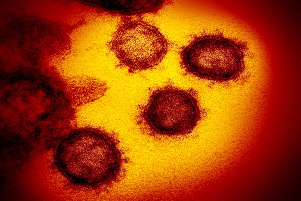
The real appearance’ of the new coronavirus: Experts in China used frozen electron microscope analysis technology to captured pictures of a strain of coronavirus after it was inactivated. This study can help in the development of drugs and vaccines against the virus.
They (researchers) also claim that the first vaccines could be in ‘clinical use’ next month
A team of researchers has released the first pictures, which show what they call ‘the real appearance’ of the novel coronavirus.
The images were captured in a lab in southern China after experts had used frozen electron microscope analysis technology to inactivate a strain of the virus.
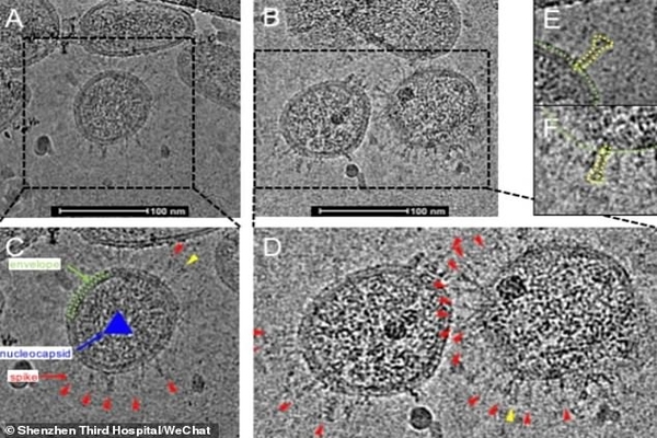
The images after using frozen electron microscope analysis technology to inactivate a strain of the virus. The pictures were released by Shenzhen Third Hospital, which is captured by the experts.
Associated professor Liu Chuang (a member of the research team), said that the appearance of the virus that we see (in the pic. above) is exactly the same as what it would be in nature.
An important intermediate state in the host cell when it was being infected by the virus, is also captured by the team
The breakthrough was jointly achieved by researchers from the Shenzhen National Clinical Medical Research Center for Infectious Diseases and Southern University of Science and Technology.
It can lay an important foundation for the identification, analysis and relevant clinical research, the team said.
Liu Lei, Party Secretary of Shenzhen Third Hospital, told Pear Video: ‘This finding will help the development of drugs and vaccines against the coronavirus.
Zhang Zheng, Director of the research institute at the Shenzhen Third Hospital, said: ‘It took us over a month. Some other places had also successfully separated [the strain of the virus].
‘But the special thing about our work is that we have not only separated [the strain], but also seen how it really looks like under the frozen microscope.’
Liu Chuang, Associated Professor from Frozen Microscopy Centre at Southern University of Science and Technology, said: ‘[The images] have a scientific significance for us to understand the life cycle of the virus.’
The team said researchers isolated a virus strain from a patient on January 27 and ‘rapidly’ completed the genome sequencing and identification.
They named the strain ‘BetaCoV/Shenzhen/SZTH-003/2020’.
Their study was published online in the pre-print journal bioRxiv yesterday.
The news comes as Chinese officials said that some vaccines for the novel coronavirus could be in clinical use next month as the number of global coronavirus cases soared past more than 100,000.